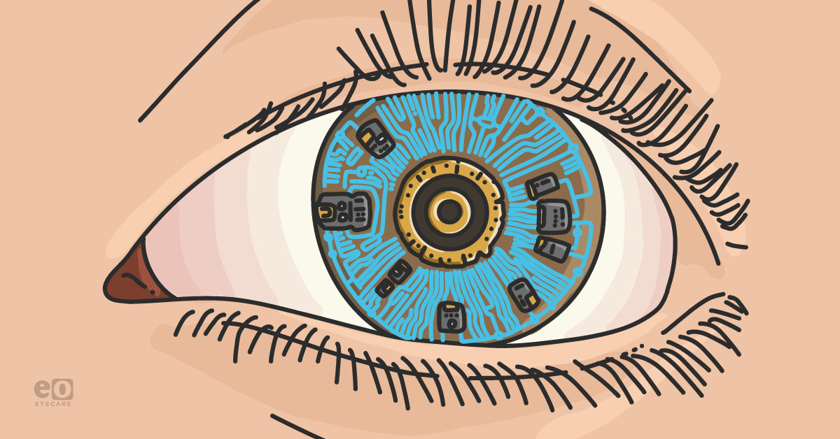
Artificial intelligence (AI) is currently one of the most popular technological trends that seems to be gaining more traction everyday. Our society’s growing appetite for instantaneous knowledge and convenience has led to the integration of AI into various facets of our daily life.
In the rapidly advancing realm of eyecare, technological innovation is being synergistically integrated with traditional medical expertise to usher in a new era of precision and efficiency. The computational capabilities of AI, machine learning (ML), and deep learning (DL) have the potential to revolutionize the diagnosis and management of most—if not all—ophthalmic conditions.
The image- and data-heavy diagnostic practices in eyecare fueled the early adoption of AI.1 Initially concentrating on the retina, researchers achieved a milestone with the US Food and Drug Administration (FDA) approval of the first fully autonomous AI diagnostic system.
The FDA-approved AI system, IDx-DR, analyzes retina photos for signs of diabetic retinopathy and determines if the patient needs to be referred to an eyecare professional or if they can be rescreened in 12 months.1
Building on this success, the heightened potential demonstrated in retinal applications has spurred increased research and focus on unlocking the potential of AI for the anterior segment of the eye.2
The differences between AI, machine learning, and deep learning
Artificial intelligence
AI is the umbrella term describing computer systems designed to mimic human intelligence by utilizing predictions and automation. AI systems are capable of performing intricate tasks such as learning, problem-solving, and decision-making.3
Machine learning
Machine learning is an advanced subset of AI that focuses on developing algorithms and statistical models to train computers to perform tasks without direct instruction. The amount of information given to the computer correlates with its ability to identify patterns and connections.
The algorithm can then utilize the pattern recognition and connections to enhance its performance, accurately anticipate outcomes, and make sound decisions when faced with new data.3
Deep learning
Deep learning is a sophisticated subset of ML designed to mimic the functionality of a human brain. DL algorithms are capable of analyzing large amounts of unstructured data to autonomously extrapolate features with less requirement of manual human intervention. All DL programs are made up of an initial input layer, followed by multiple layers of neural networks, and finalized by a terminal output layer.3
Information processed through the neural networks of a deep learning algorithm is similar to how the retina processes photons. Data is received by the input layer of the neural network and then incrementally processed and refined through several hidden layers before the end result is generated by the output layer.
Basic principles of AI algorithms in anterior segment diseases
The ophthalmic instrumentation implemented in the management of the anterior segment provides clinicians with a wealth of information. Imaging modalities such as slit-lamp photography (SLP), anterior segment optical coherence tomography (AS-OCT), Scheimpflug tomography (ST), specular microscopy (SM), and in-vivo confocal microscopy (IVCM) are each uniquely capable of capturing intricate, high-resolution images that provide in-depth analysis of various aspects of the anterior segment.
Collectively these imaging devices can provide comprehensive diagnostic and therapeutic insights. The industry-wide utilization of ophthalmic imaging devices has conveniently created a wealth of data waiting to serve as the foundational data for AI platforms designed for the analysis and interpretation of anterior segment diseases in eyecare.
Deep learning neural networks analyze pixels from images or data from labeled training sets graded by experts, learning autonomously and delivering diagnostic outputs. A thorough analysis of the dataset enables the AI algorithms to detect patterns, irregularities, and minute differences indicative of various diseases.4
However, the performance of AI and DL models is significantly influenced by the quality of the training dataset. Image annotations and grading made by ophthalmic experts must be comprehensive, accurate, and consistent to ensure an effective DL algorithm. Expansion of datasets, and incorporating diverse and complex cases can address potential biases and support the continual refinement of the algorithm.2
Using AI in the diagnosis of anterior segment diseases
AI is poised to substantially optimize the analysis of various aspects and diseases of the anterior segment.
Promising research has already exhibited the potential benefits for advancing early detection and diagnosis of diseases such as infectious keratitis (IK), diabetic peripheral neuropathy, Fuchs' endothelial dystrophy, keratoconus, dry eye disease, and cataracts. AI has also shown potential applications in improvement in the fitting and assessment of specialty contact lenses.
Infectious keratitis and artificial intelligence
The detection and management of infectious keratitis is one of the most time-sensitive diagnoses in eyecare due to the progressive and sight-threatening nature of the disease. The numerous etiologies and pathogens associated with the disease can complicate the diagnostic process. The current method of diagnosing and determining the etiology of IK involves SLP, IVCM, and tissue culture or biopsy, which is costly and time-consuming.5
The urgency of early detection and intervention of IK highlights the potential transformative role of AI. Research has been conducted to accurately detect and diagnose IK using SLP and IVCM without the traditional reliance on tissue culture or biopsy. The AI algorithms used SLP to identify structural abnormalities such as stromal infiltration, white blood cells, hypopyon, edema, and epithelial defects.
IVCM provided high-resolution images at the cellular and subcellular level that proved to be particularly useful in the diagnosis of microbial-negative IK.5 The results of these studies have shown quicker, more cost-effective results that are comparable to the accuracy of cornea specialists.
The implementation of AI in the diagnosis of IK would significantly expand the accessibility of eyecare to patients in remote areas, expediting the initiation of appropriate treatment. The scarcity of IVCM instruments in most eyecare offices is currently a limiting factor for the implementation of this technology.5
Early detection of corneal dystrophies with AI
The ability of AI algorithms to quickly analyze enormous amounts of data allows the recognition of subtle patterns that exceed the ability of even the most highly trained clinicians. This attribute proves advantageous in the detection of the nuanced changes that occur in corneal dystrophies.
Implementation of AI would not only improve the detection of corneal dystrophies, but also enhance the accuracy of early detection and predict the trajectory of the disease progression.7 As the most prevalent corneal dystrophies, Fuchs’ endothelial dystrophy (FED) and keratoconus (KCN) have been focal points of AI-related research.
Recent studies have utilized AS-OCT, ST, and SM to detect patterns in corneal thickness, endothelial cell density, the percentage of guttae, and changes in corneal topography. AI models are even demonstrating the ability to detect and diagnose subclinical manifestations of these dystrophies.7
The computing prowess of AI allows it to detect subclinical manifestations of corneal dystrophies, which can help eyecare providers offer earlier surgical intervention, minimizing any irreversible damage that could otherwise potentially occur. This technology could also be incorporated into refractive surgery consultations to detect patients with a higher risk of developing ectatic diseases even in the absence of the hallmark clinical signs that current clinicians regard as high risk.
Conversely, AI models may label some patients as suitable candidates for refractive surgery, despite traditional clinical standards classifying the patient as high risk.7
Artificial intelligence in diagnosing dry eye disease
Dry eye disease (DED) is a multifactorial disease that can lead to significant impairment of vision and quality of life if not addressed. The diagnosis and treatment of DED can be challenging due to its variety of signs and symptoms, the poor correlation between diagnostic testing and symptoms, and the low standardization of test result interpretation among clinicians.
In light of these challenges, AI has the computational power to generate intricate patterns and correlations from diverse datasets.8 AI models trained on the spectrum of clinical presentations and new DED imaging devices have the potential to establish a consensus on the diagnostic criteria for DED.
Many recent studies have focused on quantifying tear film and meibomian gland features with promising results. These advancements would enhance diagnostic accuracy and establish the foundation for more personalized and effective treatment strategies.8
The role of AI in cataract surgery
Cataracts are another area where AI is demonstrating its potential impact. With the aging trend of the population, the growing demand for cataract surgery is poised to overwhelm the eyecare system. AI has the capability to streamline the entire cataract process, from the initial diagnosis to post-operative care.9
The integration of AI in cataract care begins with the improved detection and standardized grading of the different types of cataracts. Current research has applied AI algorithms to datasets from SLP, ST, and retinal photography to quantitatively classify cataracts. This technology could be utilized in remote areas without access to specialists to provide accurate and timely cataract assessment.
AI’s impact further extends to the surgical aspect of cataracts by optimizing the consultation and surgical processes. The robust data analysis capabilities of AI can seamlessly identify patterns from datasets and compare this information to patient-specific data to formulate more individualized treatment plans and calculations.
Research is also showing potential applications for teaching, training, and real-time feedback during cataract surgery to improve surgical decision-making and reduce complication potential.
Fitting specialty contact lenses with AI
Contact lenses, initially created for the purpose of independence from glasses, have evolved to address other eyecare needs as well. Modern contact lenses are being utilized as effective tools for myopia control and dry eye disease. Research is also being conducted to utilize contact lenses as a drug delivery device or biosensor for monitoring real-time physiological parameters.10
The fitting of specialty contact lenses can be challenging due to the unique and varying anatomical structures of each eye, leading to multiple office visits and extensive chair time. Implementing AI models with detailed objective measurements from SLP, AS-OCT, and ST could identify patterns that yield a more accurate empirical contact lens fitting and also guide any needed contact lens modifications.
Increasing the efficiency and accuracy of specialty contact lens fittings could enhance the overall experience for the patient and the clinician.10 While the development of drug delivery and biometric monitoring contact lenses still seems to be in the early stages, the benefits of AI are already being considered.
AI models could regulate the release of drugs from contact lenses based on real-time changes detected through biometric monitoring. This technology would significantly increase patient compliance and treatment efficacy of some ocular diseases such as glaucoma.10
Current limitations of artificial intelligence in eyecare
The integration of AI into the ophthalmic space has seemingly endless potential, most notably in improving the objectivity and consistency of diagnostics. The majority of the ophthalmic AI systems that exist are still in developmental stages and are only applied in a research setting.
While the potential of AI in the ophthalmic space seems immense, the limitations and challenges that must be addressed before clinical application cannot be ignored.2
Data standardization and annotation
Data standardization is among the primary challenges of AI implementation in the clinical eyecare space. The numerous anterior segment ophthalmic imaging platforms generate data with variations in magnification, contrast, and image resolution, making the standardization of images for AI algorithms extremely difficult.
Consistency in the quality and grading of the images would also be necessary to ensure the accuracy of the algorithm.2 Most ophthalmic AI algorithms that exist are ML models, requiring clinical experts to annotate and grade images before they can be added to a training data set.
The task of data annotation is time-consuming, and the ML models require large data sets to ensure accuracy, placing a significant burden on the experts constructing the data sets.2
Explanations for AI-based diagnoses
The decision-making process by which an AI algorithm arrives at its diagnosis is not always apparent to clinicians, creating somewhat of a black-box phenomenon.
With the responsibility of the diagnosis falling on the clinician, trusting a diagnosis without a clear understanding of the reasoning can be unsettling. Explainable AI models are being developed to alleviate this concerning obstacle.2
Diagnostic errors in AI
The most worrisome aspect of AI integration into eyecare is the possibility of diagnostic error and the potential for disastrous effects on someone’s vision or overall health. A recent study investigated the reliability and accuracy of ChatGPT’s ability to provide information regarding various eye diseases and their appropriate management.
A grading system was established based on guidelines from the American Academy of Ophthalmology. While most responses were considered accurate, a significant amount of responses were also missing pertinent information, and a small portion (7.5%) of responses were deemed potentially dangerous.11
Rigorous standards and continuous monitoring must be implemented to minimize the risks associated with diagnostic errors and prioritize patient safety. Despite the apparent positive impact of AI in eyecare, the collective acceptance among clinicians and patients will be a gradual process.2 The doctor-patient relationship and individual patient needs are additional aspects of health care that are unlikely to be fully replaced or replicated by AI systems.
In conclusion
The fusion of eyecare and AI has great potential to enhance diagnostic accuracy and patient outcomes. Early disease detection, prediction of disease progression, and tailored treatment plans are all within the computing capabilities of AI algorithms. The growing demand for high-quality medical services, efficiency in healthcare delivery, and advancements in technology are driving researchers to maximize the AI potential in many aspects of healthcare.12
While AI will likely become a commonplace tool to elevate our standards of care, many challenges will need to be addressed before implementation into direct patient care. Additionally, human interaction and emotion that reinforces relationships between doctors and patients is vital to providing individualized care, and something that AI cannot replicate.
The irreplaceable value of human interaction and trust between doctors and patients underscores the importance of integrating AI as a complementary tool rather than a replacement for healthcare providers.
That being said, it is important to recognize that while AI will likely become a routine and powerful tool that will elevate our standards of care, there are still current challenges that need to be overcome before this occurs. Additionally, the use of AI does not replace the relationship between doctors and patients, and the need for individualized care rooted in both clinical data and personal empathy will remain vital to providing comprehensive patient care.
